RETINAL DETACHMENT
What is retinal detachment?
Retinal detachment is an ocular disorder that occurs due to the spontaneous separation of the neurosensory retina (inner layer of the retina) from the pigmentary epithelium (outer layer).
It is a serious visual problem that can occur at any age, although it usually occurs in middle-aged individuals or elderly people. It is usually more frequent in people who are myopic or in those who previously had a retinal disorder.
It is very important to make a diagnosis as quickly as possible, since the chances of improvement are greater if the macula or central area of the retina is not affected.
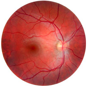
Normal retina image
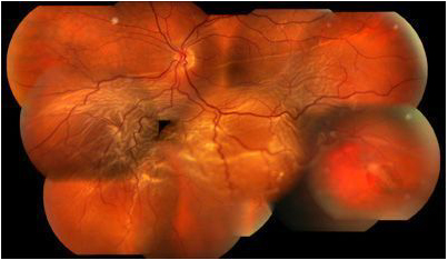
Retinal detachment image where the tears are clearly visible
Symptoms of Retinal Detachment
- Sudden appearance of floaters or “flying flies” or an increase in existing ones.
- Appearance of flashing lights
- Appearance of a shadow on one side of the visual field
- Appearance of a grey curtain moving in the middle of the field of vision
- A sudden decrease in vision.

Treatment
Laser and Cryotherapy: Most retinal tears should be treated by fixing the retina to the posterior wall of the eye. Both laser (photocoagulation) and cryotherapy (freezing treatment) create a scar that helps fix the retina, preventing the passage of fluid through the tear, preventing the retina from detaching. When retinal tears exist, it is advisable to perform a preventive treatment, usually by laser, even though they have not yet caused a detachment. This preventive treatment can also be useful for high risk patients with peripheral degenerative lesions that can end in a break.
Surgery: Almost all patients with retinal detachment have to undergo a surgical repositioning of the retina. The method to fix the retina depends on the characteristics of the detachment. It can be approached through scleral surgery where a buckle (ring or cerclage) is applied to the outermost wall of the eye to counteract the traction forces on the retina and help it to approach its normal position, or perform a vitrectomy, a surgical technique where the vitreous (the gel that fills the ocular cavity) is extracted in order to work directly on the retina and then apply it again. This last surgical technique is used for several vitreo-retinal pathologies.

Vitrectomy technique